Hollow Gold Nanoshells for Sensitive 2D Plasmonic Sensors
Daoming Sun (1,2), Ferdaous Ben Romdhane (3), Michèle Salmain (2) and Souhir Boujday (1)
Daoming Sun 1,2, Ferdaous Ben Romdhane3, Michèle Salmain 3, Souhir Boujday1
1Sorbonne Université, CNRS, Laboratoire de Réactivité de Surface (LRS), 4 place Jussieu, F-75005 Paris, France
2Sorbonne Université, CNRS, Institut Parisien de Chimie Moléculaire (IPCM), 4 place Jussieu F-75005 Paris, France
3Sorbonne Université, CNRS, FCMat, Campus Pierre et Marie Curie, 4 Place Jussieu, F-75005 Paris Cedex 05, France
The interaction of incident light with noble metal nanoparticles engenders a fascinating phenomenon known as localized surface plasmon resonance (LSPR). This results in the presence of single or multiple intense absorption bands in the visible to near-infrared spectral range whose position is affected by the refractive index of the surrounding medium. In this comprehensive study, we thoroughly investigated the experimental parameters governing the size, aspect ratio, and optical properties of hollow gold nanoshells (hAuNS) synthesized through galvanic exchange of cobalt-based nanospheres. Subsequently, we rigorously determined both the empirical and theoretical refractive index sensitivity (RIS) and figure of merit (FoM) of these engineered nanostructures. Notably, hAuNS with an external diameter of 98 nm and a shell thickness of 13 nm demonstrated a noteworthy RIS of 360 nm/RIU and a FoM of 2.0 in solution. In contrast, solid gold nanospheres (sAuNS) of a similar diameter exhibited a significantly lower RIS of 136 nm/RIU. Following the transfer of both these nanostructures onto glass slides for the development of LSPR sensors, it was intriguing to note that the RIS and FoM remained largely unaffected. These findings underscore the potential of these plasmonic nanoparticles as promising candidates for the design of sensitive solid-phase LSPR sensing devices
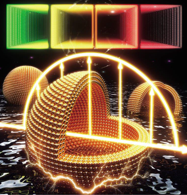
Hollow Au nanoshells dispersed in water show electro-optical effects due to the LSPR phenomenon. The center and right particles were dissected and partially immersed in water to reveal their hollow, solvent-filled interiors. The upper squares represent the LSPR band range, from green light to NIR